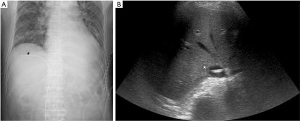
A man in his 70s was admitted to our intensive care unit after neck surgery. An ultrasound-guided right femoral central venous catheter (CVC) was introduced, without difficulty. We didn’t use a subclaviclar catheter because the operator was concerned about venous stenosis and congestion. The position of the tip of the catheter from the puncture point was 50 cm. A chest X-ray showed an anomalous bend of the CVC with the tip below the right diaphragm (Figure 1A). An echography showed the CVC placed (asterisk) in the inferior vena cava (IVC) (Figure 1B). The IVC showed short-axis view, but the CVC didn’t show short-axis view. This was indicated that the CVC was not straight up within the IVC. Radiographic and echographic findings suggested that the catheter entered the hepatic vein through the IVC. The view of the CVC within a hepatic vein was very poor and not recorded.